Descriptive Study of Gastroduodenal Lesions in the Internal Medicine Department of Point G University Hospital
Author'(s): Traoré Djenebou1,2*, Sangaré Moussa1, Keita Zakaria3, Sy Djibril1,2, Boa Ange Trévis1, Sinayoko Adama1, Goita Issa Souleyname2, Koné Nouhoum1, Nyanke Nounga Romuald1, Landouré Sekou1, Keita Kaly1, Mallé Mamadou1, Cissoko Mamadou1, Dembélé Ibrahim A1, Fané Sékou1, Diarra Aoua1, Koné Yacouba1, Dao Karim4, Sangaré Drissa4,5, Tolo Nagou6, Togo Mamadou6,4, Traoré Abdramane6, Mahamadou Saliou4,6, Camara Boua Daoud7, Soukho Assetou Kaya1,2
1Internal Medicine Department of CHU Point G of Bamako.
2Faculty of Medicine and Odontostomatology of Bamako.
3University Clinical Research Center (UCRC).
4Internal Medicine Department of CHU Gabriel Touré of Bamako.
5Internal Medicine Department of Fousseyni Daou Hospital in Kayes.
6Internal Medicine Department of CHU Boubacar Sidi Sall of Kati.
7Internal Medicine Department of Fousseyni Daou Hospital in Segou.
*Correspondence:
Djénébou Traoré, Internal Medicine Department of CHU Point G. Tel: 00222376466129 / 66466129.
Received: 15 Jun 2024 Accepted: 28 Jul 2024
Citation: Traoré Djenebou, Sangaré Moussa, Keita Zakaria, et al. Descriptive Study of Gastroduodenal Lesions in the Internal Medicine Department of Point G University Hospital. Gastroint Hepatol Dig Dis. 2024; 7(4): 1-4.
Abstract
Introduction: The practice of biopsies during digestive endoscopy is a multi-daily procedure in gastroenterology and gives this endoscopic examination a dual objective, first macroscopic by the description of the lesions and their architecture and then by taking samples for analysis histopathological. The study aimed to determine macroscopic and histological lesions in gastric and duodenal biopsies.
Methodology: This was a cross-sectional and analytical study over 12 months from June 15, 2021, to June 15, 2022, in patients who had undergone esophagus -gastro-duodenal fibroscopy (FOGD) with biopsy.
Result: The study included 165/378 patients who underwent an anatomopathological examination with a search for H. pylori after gastric biopsy, a frequency of 43.65%. Of these, 133/165 or 80.61% had H pylori in their samples. The sex ratio was 0.87. The mean age was 47.5 years ± 14.81 years. Among the clinical information that motivated the performance of the FOGD, epigastralgia predominated with 80.0%. The endoscopic lesion topography was gastric in 90.3%. The type of lesion was erythematous and erosive gastritis in 43.03%. The histological appearance of the lesion was chronic gastritis in 86.66% followed by gastric tumor in 11.5%. Among these gastric tumors, 77.78% were adenocarcinomas. The more H pylori was present, the more patients had a risk of having chronic gastritis (p = 0.005 with a RR = 12.33) CI [5.16-29.46].
Conclusion: It appears from our study that H. pylori infection was associated with all gastroduodenal pathologies with a higher frequency in benign ulcers at 92.9% and in chronic gastritis at 89.51% which was the most encountered histological aspect; Gastric adenocarcinoma, which constitutes a public health problem in Mali was also strongly associated with H. pylori at 80.0%.
Keywords
Introduction
Digestive endoscopy allows the exploration of the digestive tract, the bile ducts and the pancreas, and most often allows the performance of a therapeutic procedure. It is the reference examination for the diagnosis of pathologies of the esophagus- gastro-duodenal mucosa: esophagitis, oesophagus varices, cancers of the stomach and oesophagus, peptic ulcer, etc. [1].
Endoscopy has become the reference examination of the esophagus-gastro-duodenal mucosa due to its good tolerance, speed, possibility of biopsy, and even of having therapeutic action. It has replaced the esophagus-gastro-duodenal transit which is no longer indicated by the College of Radiology Teachers of France [1].
The practice of biopsies during digestive endoscopy is a multi- daily procedure in gastroenterology and gives this endoscopic examination a dual objective, firstly macroscopically by describing the lesions and their architecture and then by taking samples for histopathological analysis. Biopsies are essential for the diagnosis, monitoring and treatment of certain pathologies [2].
During gastroscopy: systematic gastric biopsies can be used to detect H. pylori infection and preneoplastic lesions. It also makes it possible to carry out the bacteriological examination with evaluation of the sensitivity of the bacteria to antibiotics, if they are available [3].
In the absence of contraindications to biopsies, it is recommended to perform:
- For the pathological examination: at least 5 gastric biopsies (2 at the antrum level, 1 at the angle level and 2 at the body level) for the diagnosis of the infection (preferably by immunohistochemistry) and study inflammatory activity, atrophy and intestinal metaplasia (new OLGA1 or OLGIM2 classifications making it possible to assess the risk of progression to gastric cancer depending on the degree of extent and severity of preneoplastic lesions).
- And for the bacteriological examination with assessment of sensitivity to antibiotics, if feasible: 2 additional biopsies (at the antrum and body) to be sent to the bacteriology laboratory in a specific transport environment [3].
Thus, to study the profile of the results from the biopsy of the gastric mucosa in Mali, we initiated this study in the internal medicine department of the University Hospital Center (CHU) of Point G. This will allow us to determine the frequency by anatomy- pathology of the gastric mucosa diseases.
Patients and Methods
We carried out a cross-sectional and analytical study with the prospective collection over 12 months from June 15, 2021, to June 15, 2022, in patients who performed an FOGD with biopsy in the internal medicine department whose tissue sample was sent to the department of the anatomy-pathology of Point G University Hospital for histological analysis associated with the search for Helicobacter pylori. The variables studied were socio-demographic, clinical, biological, endoscopic and anatomopathological aspects. Data entry and analysis were carried out using SPSS software.
Results
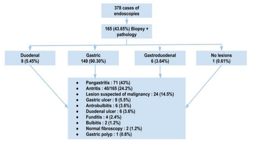
The study included 165/378 patients who underwent a pathological examination with a search for H. pylori after gastric biopsy.
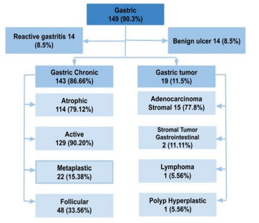
H.pylori-positive patients represented 80.61% of our sample (133/165). The sex ratio was 0.87. The average age of the patients was 47.5 years ± 14.81 years, with extremes of 14 years and 83 years. Urban origin (Bamako) was the majority with 73.30% of cases. Housewives predominated in our study at 33.33%. The dominant clinical sign, motivating the performance of FOGD, was epigastralgia (80.0% of cases). The endoscopic lesion topography was gastric in 90.3%. The type of lesion was erythematous and erosive gastritis in 43.03% followed by arthritis and lesions suspicious for malignancy in 24.24% and 14.55% respectively. The predominant histological appearance was chronic gastritis in 86.66% followed by gastric tumor in 11.5%. Chronic gastritis was active in 90.20% of cases. Among these gastric tumors, 77.78% were adenocarcinomas. The more H pylori were present, the more patients had a risk of having chronic gastritis (p=0.005 with an RR=12.33) CI [5.16-29.46].

Figure 1: Distribution of macroscopic, topographic and lesional aspects according to upper digestive endoscopy.

Figure 2: Distribution of gastric lesions on a histological basis.
Discussion
On the macroscopic level, according to the lesion topography in upper digestive endoscopy; the lesions predominated at the gastric level (90.30%). Tolo [4] made the same observation 80.99%. This would perhaps be linked to the socio-demographic characteristics of our study populations, given that all these studies concerned the same demographic sphere with different study objectives.
Our overall H.pylori frequency was 80.61% in gastric and duodenal biopsies; a result comparable to numerous studies carried out on the subject; notably that of Konaté in Mali with 89.4% [6], Dia in Senegal with 78.5% [7] and Somda in Morocco with 83.76% [8]. But not superimposable to the prevalence of H. pylori in industrialized countries which was 30 to 52% according to Zeitoun [1]. This difference could be explained by the fact that in our context, socio-sanitary conditions are often precarious, and favourable to the transmission of the germ.
The female gender represented 53.33% with a sex ratio of 0.875; on the professional level we found a predominance of housewives i.e. 33.33%. This same observation was made by Ngouala G [10] in Senegal which also found not only a clear predominance of feminism (including 175 women and 73 men, i.e. a sex ratio of 2.3) but also on the professional level a dominance of women in the field household, i.e. 55% of cases. Its data can be explained by the fact that in Africa in general, especially the West African side, housekeeping is a profession too often reserved for women. Households in Africa are generally exposed to enormous chemical compounds (wood and coal smoke, dust, etc.) which constitute exogenous factors known as environmental factors which can constitute a means of weakening feminism's health about men.
The patients in our study were relatively young with a mean age of 47.5 ± 14.81 years, and ranges of 14 and 83 years. Similar results were observed in Senegal by Dia (48.7 ± 17.45 years) [7], in Togo by Lawson-Ananissoh (43.56 ± 18.54 years) [11].
Epigastralgia (80.0%) was our main symptom motivating the performance of oeso-gastro-duodenal fibroscopy. This result is comparable to that of Youssouf [12] who found epigastralgia and weight loss as the main symptom with 91.9% each in a study focused on stomach neoplasia. At endoscopy, the most common macroscopic lesion found was pangastritis at 43%. Tolo [4] mainly observed gastritis in 50.0% of cases. The acute phase of H. pylori infection gives a focal inflammatory attack that extends to the entire gastric mucosa with the persistence of the germ, we witness its chronic evolution.
The predominant histological type was chronic gastritis in almost 88.66% of cases; this chronic gastritis was active in 90.20% and atrophic in 79.72%. It is associated with follicular and metaplastic gastritis in 33.56% and 15.38%. These data are close to that of Tolo [4] who found a proportion of 86.96% of cases of chronic gastric. The history of the natural evolution of the damage to the gastritis mucosa can explain this correlation. We found a gastric tumor in 19 patients after anatomopathological examination, i.e. 11.5%. Among our collected histological tumor types, we observed a clear dominance of adenocarcinoma (77.78%); followed by gastrointestinal stromal tumor (11.11%) and gastric lymphoma as well as a hyperplastic gastric polyp in the same way, i.e. 5.58% each. The result is different from that of Youssouf [12] which, apart from the predominance of adenocarcinoma as the majority histological type (91.9%); also identified other histological types such as sarcoma and squamous cell carcinoma with respectively 5.4% versus 2.7%. This correlation of the histological type of adenocarcinoma could be explained by the fact that it is the most common histological type of gastric cancer.
H.pylori infection was associated with all gastroduodenal pathologies with a higher frequency in benign ulcers at 92.9% and in chronic gastritis at 89.51% which was the most common histological appearance. Gastric adenocarcinoma was also strongly associated with H. pylori at 80.0% and constitutes a public health problem in Mali.
Conclusion
It appears from our study that H. pylori infection was associated with all gastroduodenal pathologies with a higher frequency in benign ulcers at 92.9% and in chronic gastritis at 89.51% which was the most common histological appearance; Gastric adenocarcinoma which constitutes a public health problem in Mali was also strongly associated with H. pylori at 80.0%.
References
- Zeitoun JD, Chryssostalis A, Lefevre Hepatology gastroenterology visceral surgery. 7th edition, Paris: Vernazobres-Grego. 2020; 609.
- Chabrun E. Biopsies in digestive endoscopy: good practice POST’U. 2021; 10: 247-256.
- Haute Autorité de Santé (HAS) (RELEVANCE OF CARE - Diagnosis of Helicobacter pylori infection in adults), National Professional Council of Hepato-Gastroenterology. 2017;
- Tolo N, Binan Y, Apeti S, et Esogastroduodenal fibroscopy in the elderly in Bamako: A study of 121 cases. Health Sci Dis. 2023; 24: 157-161.
- Mbengue M, Boye CS. Helicobacter pylori in Africa. Acta 1999; 29: 525-526.
- Holcombe C, Omotara BA, Eldridge DJ, et al. Helicobacter pylori the most common bacterial infection in Africa: a random serological study. Am J Gastroenterol. 1992; 87: 28-
- Konaté A, Diarra M, Soucko-Diarra A, et al. Chronic gastritis in the era of Helicobacter pylori in Mali. Acta Endoscopica. 2007; 37: 315-320.
- Dia D, Seck A, Mbengue M, et al. Helicobacter pylori and gastroduodenal pathology in Dakar (Senegal). Med 2010; 70: 367-370.
- Somda SK, Coulibaly A, Koulidiati J, et al. Helicobacter pylori infection and gastroduodenal pathologies at the Ibn Sina University Hospital Center in Science and technology, health science. 2016; 39: 83-89.
- Ngouala GABB, Bourgi L, Da Veiga JAI, et Upper digestive endoscopy in Louga (Senegal): patient profile and difficulties encountered. PAMJ. 2017; 27: 211-216.
- Lawson-Ananissoh LM, Bouglouga O, Bagny A, et Epidemiological profile of peptic ulcers at the Lomé Campus hospital and university center (TOGO). African Journal of Hepato Gastroenterology. 2016; 9: 99-103.
- Youssouf O, Diarra M, Samake K, et al. Epidemiological, Clinical and Histological Aspects of Stomach Cancer at the Gabriel Touré University Hospital of Bamako (Mali). ESJ. 2022; 18: 123-130.
- Koura M, Ouattara DZ, Some RO, et Chronic gastritis at the Sourô Sanou University Hospital Center in Bobo-Dioulasso (Burkina Faso): epidemiological, clinical, endoscopic and histological aspects. AJOL. 2020; 43: 124-134.