Paget's Disease: Striking Clinical Findings and With New Palliative Topical Therapy Using 25% Podophyllin
Author'(s):Khalifa E. Sharquie1 and Wesal K. Al-Janabi2
1Department of Dermatology, College of Medicine, University of Baghdad, Iraqi and Arab Board for Dermatology & Venereology,Baghdad Teaching Hospital, Medical City, Baghdad, Iraq.
2Dermatology Center, Medical City, Baghdad, Iraq.
*Correspondence:
Dr. Wesal Aljanabi, Dermatology Center, Medical City, Baghdad,Iraq.
Received: 18 August 2019; Accepted: 17 September 2019
Citation: Khalifa E. Sharquie, Wesal K. Al-Janabi. Paget’s Disease: Striking Clinical Findings and With New Palliative Topical Therapy Using 25% Podophyllin. Cancer Sci Res. 2019; 2(2); 1-6.
Abstract
Background: Paget’s disease is a rare cancer whether mammary or extra-mammary but must be considered in any persistent eczematous eruption of nipple &/ areola or elsewhere like genitalia which does not responds to topical treatment. Although it is slowly growing tumor but diagnosis and therapy should be carried out before deep invasiveness might occur.
Objective: To report cases of Paget’s disease in order to pay attention to the increasing frequency of this rare disease among population especially among females.
Patients and Methods: This study was conducted at the Dermatology Center, Medical City, Baghdad, Iraq, during the period from May 2007 to Oct. 2019. All the cases of biopsy proven Paget’s disease were evaluated for the demographic data like age, gender, family history. In addition, clinical features such as site, underlying palpable breast mass, regional lymphadenopathy, examination of contralateral breast and any associated systemic symptoms. Relevant work up to find any associated underlying malignancy was done. A number of patients who refused surgery were treated by topical podophyllin 25% solution at weekly sessions for minimum six weeks with follow up period of one year.
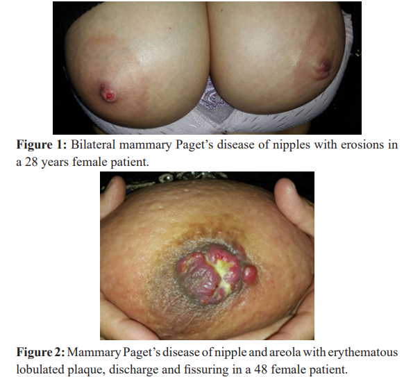
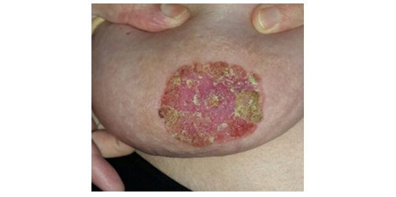
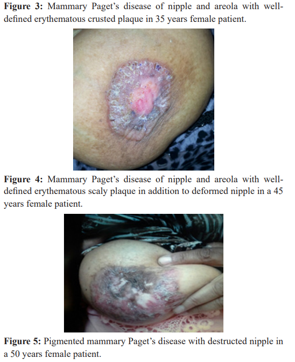
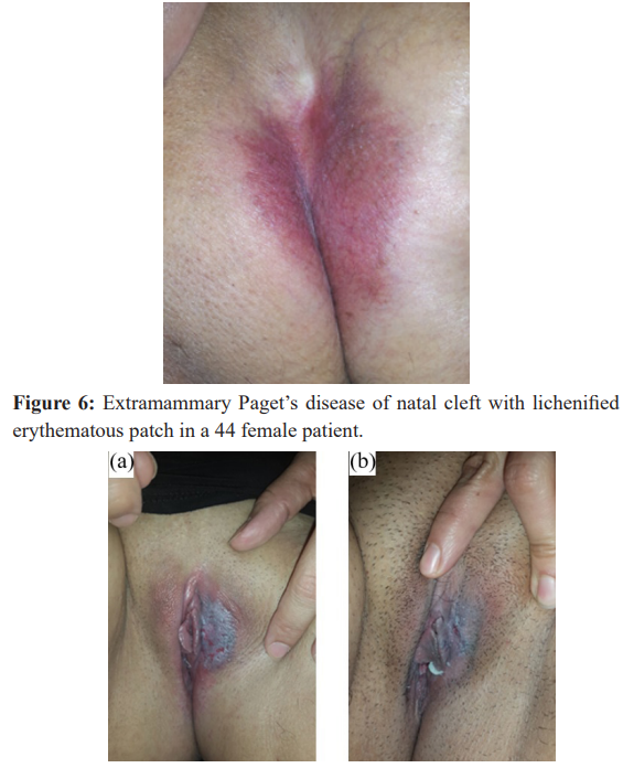
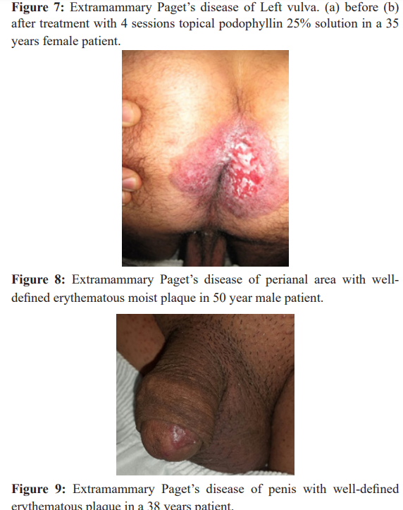
Results: Twenty-One patients were included in this study, 19 females and 2 males (9.5:1). The mean age was 40.2 ± 5.3 (range 28-53) years. The duration of lesions ranged between 2 months -1 year. Sixteen (76.19%) cases were mammary Paget's disease (MPD), while 5(23.8%) cases were extramammary (EMPD). The frequency of MPD in relation to EMPD was 3.2:1. Regarding MPD, the right breast was involved in 12 cases, while only 2 cases affecting the left breast. Two cases (12.5%) had bilateral MPD (1 nipples only and the other nipples and areolas). Ten (62.5%) cases affected the nipple and areola together (one case bilateral), whereas in 6 (37.5%) cases the nipple (one case bilateral) was only diseased. The lesions of nipple and/areola presented as erythematous, scaly, crusted, some with oozing, erosions and even ulcerations. The nipple looked atrophied to nearly eroded in 3 (14.82) cases. Pigmented MPD was seen in 4 (25%) patients. For the five patients with EMPD, 2 female patients had natal cleft lesions (1 with erythematous patch and the other with hypopigmented patch) and one patient with left vulvar lesion. Perianal erythematous moist eroded plaque was seen in one male patient while another male patient had penile erythematous plaque. All the lesions of both MPD and EMPD were well defined. The associated symptoms were mainly itching, burning and discomfort No underlying palpable breast mass (MPD) or associated underlying internal Or malignancy (EMPD) and no regional lymphadenopathy nor distant metastasis were detected. Topical podophyllin was applied in 7 cases, six on breast and one vulvar who refused surgery and it had good palliative action preventing progressing their lesion.
Conclusion: We think that Paget's disease is increasing among Iraqi females when compared with previous years. Paget’s disease must be kept in mind when facing persistent eczematous involvement of the nipple and/ areola, or elsewhere not responding to a local treatment as urgent intervention is highly recommended. Topical therapy (like using topical podophyllin) is an option for isolated disease and as an adjuvant in patients who refused radical treatment.
Keywords
Introduction
Paget’s disease (PD) is an uncommon intraepithelial adenocarcinoma occurring in apocrine bearing skin [1]. Mammary Paget’s disease (MPD) of nipple and/ areola represents 1-3% of breast cancers. The peak incidence is between 50 and 60 years of age, mostly in women. It is isolated on 1.4-13.3% of patients. In various studies, 82-100% of cases were associated with either in situ (13.3-52%) or invasive (30-60%) ductal carcinoma [2,3].
Extramammary Paget’s disease (EMPD) is a rare malignancy of the anogenital region, occurring mostly in the 6th through 8th decades of life, more in women [4-6]. EMPD is associated with an underlying adnexal carcinoma in or visceral malignancy in approximately 20-30% of cases [5], such as bladder, urethra, prostate and colorectal cancers [7]. In rare reports, ectopic EMPD had been reported in areas that are relatively free of apocrine glands, such as the chest, abdomen, thigh, eyelids, face, and external auditory canal [1,8].
Both MPD and EMPD present with a long-standing history of erythematous, scaly or velvety patches or plaques on breast and anogenital skin respectively. Because of the rather nondescript appearance, there is often a several month- delay in diagnosis as a presumed inflammatory or infectious dermatitis. MP usually involve the nipple and / areola with ulceration, weeping and crusting are often present. Nipple erosion and discharge may occur. The associated pruritus may lead to lichenifications and excoriations. Retraction of the nipple and areola can occur in advanced cases. In EMPD, hypo- and hyper pigmentation may also occur [1].
Historically, two contrasting theories attempted to explain the pathogenesis of MPD: The first is the epidermotropism (currently favored in literature) in which malignant Paget’s cells arise from an underlying breast adenocarcinoma and directly extend into the epidermis via the lactiferous ducts. Immunohistochemically studies showed significant similarities between Paget’s cells and the underlying breast carcinoma in a majority of cases. In contrast, epidermal keratinocytes expressed a different Immunohistochemically staining pattern. The second theory is the transformation of epidermal keratinocytes or degeneration into malignant Paget’s cells [1,9]. EMPD represents malignant cells that are believed to originate from the intraepidermal parts of apocrine glands or from pluripotent cells in the epidermis [1].
One half of MPD were found concurrently to have a palpable underlying breast mass [10]. Of these, one half to two thirds have axillary lymph node metastasis [10,11]. Therefore, for MPD bilateral mammography is required in all cases and MRI has increased sensitivity [1,12]. For EMPD, colonoscopy, cystoscopy and in female patient, pelvic examination with Papanicolaou test and colposcopy, should all be considered as first line studies. Other imaging studies can be considered accordingly such as pelvic ultrasound, MRI, mammography, and intravenous pyelogram [13].
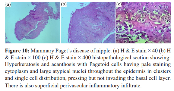
Punch, wedge or excisional biopsy of lesional skin is necessary to confirm the diagnosis [1]. The intraepidermal adenocarcinoma of EMPD and MPD has a similar histologic appearance. There are groups, clusters or single cells within the epidermis that show nuclear enlargement with atypia, prominent nucleoli and ample cytoplasm [14]. The cells may be in all levels of epidermis, and compress, but preserve the basal layer without junctional nest. The cells may extend into the contiguous epithelium of hair follicles and sweat bland ducts. Acanthosis, hyperkeratosis, and parakeratosis are often present. These cells have pagetoid appearance and simulate other intraepidermal malignancies, such as melanoma, pagetoid squamous cell carcinoma, mycosis fungoides, cutaneous adnexal carcinomas (e.g., sebaceous carcinoma and porocarcinoma), Merkel cell carcinoma and Langerhans cell histiocytosis. The cells stain positive for PAS stain, mucicarmin, Alcian blue stain and colloidal iron and are diastase resistant [15].
Patients and Methods
This study was conducted at the Dermatology Center, Medical City, Baghdad, Iraq, during the period from May 2007 to Oct 2019 that was conducted by one doctor (KE Sharquie). All the cases of pathologically proven Paget’s disease were evaluated for the demographic data like age, gender, family history. In addition, clinical features as site, underlying palpable breast mass, regional lymphadenopathy, examination of contralateral breast and any associated systemic symptoms. Relevant work up to find any associated underlying malignancy. Topical podophyllin was applied in 7 (33.33%) cases that refused radical intervention like surgery, six on breast and one vulvar. Topical Podophyllin 25% was prepared by mixing 25g of Podophyllum resin in 100 cc tincture benzoin. This therapy was applied once a week for minimum six weeks. Pre, during and post treatment photographs were taken for efficacy evaluation and follow up. Data entry followed by inferential statistics was carried out by the researchers using the software statistical package for social sciences (SPSS) version 20. Continuous variables presented as mean ± SD (Standard deviation) and discrete variables as numbers and percentages.
Results
Twenty-One patients were included in this study, 19 females and 2 males (9.5:1). The mean age was 40.2 ± 5.3 (range 28-53) years. The duration of lesions ranged between 2 months -1 year. Sixteen (76.19%) cases were mammary Paget's (MPD) (Figure 1-5), while 5(23.8%) cases were extramammary (EMPD) (Figures 6-9). The frequency of MPD in relation to EMPD was 3.2:1. Regarding MPD, the right breast was involved in 12 cases, while only 2 cases affecting the left breast. Two cases (12.5%) had bilateral MPD (1 nipples only and the other nipples and areolas) figure1. Ten (62.5%) cases affected the nipple and areola together (one case bilateral), whereas in 6 (37.5%) cases the nipple (one case bilateral) was only diseased. The lesions of nipple and/areola presented as erythematous, scaly, crusted, some with oozing, erosions and even ulcerations (Figure 2-4). The nipple looked atrophied to nearly eroded in 3 (14.82) cases (Figure 3-5). Pigmented MPD was seen in 4 (25%) patients. For the five patients with EMPD, 2 female patients had natal cleft lesions (1 with erythematous patch (Figure 6), and the other with hypopigmented patch) and one patient with left vulvar lesion (Figure 7). Perianal erythematous moist eroded plaque was seen in one male patient, figure 8, while another male patient had penile erythematous plaque (Figure 9). All the lesions of both MPD and EMPD were well defined. The associated symptoms were mainly itching, burning and discomfort No underlying palpable breast mass (MPD) or associated underlying internal malignancy (EMPD) and no regional lymphadenopathy nor distant metastasis were detected. Topical podophyllin had good palliative action as the lesions regressed with improvement in ulceration but one, the vulvar case, had progressed after one year with extensive local metastasis.
Discussion
Paget’s disease is carcinoma in situ that might involve any area in the body but commonly the nipple and genitalia. Paget’s disease in Iraqi population was rarely seen before 1990, but since then, the cases are increasing and commonly involving the nipple and sometimes the genito-anal area, often miss diagnosed as discoid dermatitis especially when unilateral. This increase in its frequency go parallel with increase with other malignancy like Kaposi Sarcoma [16], Malignant Melanoma [17], Dermatofibrosarcoma protuberance [18] and Mycosis Fungoides [19] basal and squamous cell carcinoma [20]. In the present study, the age at presentation (40.2 ± 5.3years) was younger than reported in literatures (above 50 years) [2,3]. The frequency of MPD in relation to EMPD was 3.2:1 in the present work. Women were found to outnumber (9.5:1) men in this study and this is comparable to what had been described in publications [1,2]. The nipple and areola were more commonly affected than nipple alone in this study. Bilateral MPD was present in only 2 cases (12.5%), as rare [21,22], as about only 13 cases of bilateral MPD had been reported [23]. Pigmented MPD was seen in 4(25%) of this work cases, with only about 14 cases of pigmented MPD had previously been reported [24,25]. In addition, it has been reported that EMPD had underlying adnexal carcinoma or visceral malignancy in approximately 20-30%, but in the present study no underlying cancer was discovered [5].
Evidence based treatment recommendations for MPD are not available because of lack of studies directly comparing outcomes for mastectomy versus breast conservative therapy (BCT) followed by radiotherapy [1]. Sentinel lymph node biopsy (SLNB) is recommended for all patients [26].
For EMPD, invasive disease has a greater recurrence rate than in situ disease (67% vs 35%) following surgical, even with wide local, excision Margins [5,27]. Some adjuvant techniques had been used to decrease the recurrence, such as intraoperative frozen section analysis, [28], multiple scouting biopsies [29], and intraoperative CK7 immunostaining during Mohs microscopic surgery [30]. Some advocate the use of SLNB and/ regional lymph node dissection in all high-risk EMPD cases, despite negative findings on PET-CT, as micrometastasis are undetectable [31]. Radiotherapy has been used as a primary treatment [32], and after surgery [33], or as an adjuvant therapy in high-risk group [34]. No randomized controlled studies comparing surgery to radiotherapy have been performed yet [1]. 5-FU cream has been used as an adjuvant modality prior to surgery and for early post-operative detection of recurrence [35,36]. However, 5-FU has not proven to be a reliably curative agent in the treatment of EMPD because of the limited penetration of the drug and the inability to reach the deeper epidermal layers and adnexal structures [36]. Immiquimod has been reported to result in clinical and histologic clearance in a few case series involving primary and recurrent EMPD; some of the later declined further surgery for their 2nd recurrences and chose to be retreated with [37]. Systemic chemotherapy has been used in cases of invasive and metastatic disease or when surgery and radiotherapy are contraindicated [1]. The prognosis depends on the presence of a palpable mass and the invasiveness of the underlying cancer [38].
Podophyllin is an alcoholic plant extract obtained from dried rhizomes of common plants called emodi (Indian Podophyllum) or Podophyllum peltatum (May apple or Mandrake). Podophyllum resin contains many active ingredients that work through multiple actions mainly by arresting mitosis and differentiation, blocking oxidation enzymes in Tricarboxylic acid cycle and thus interfering with cell nutrition, inhibiting axonal transport, protein, RNA, and DNA synthesis and affecting mitochondrial activity with reducing cytochrome oxidase activity. Podophyllin had been used over a long time for the treatment of viral warts, Condyloma Acuminatum with report of use in genital warts in infants, oral hairy leukoplakia, juvenile papilloma of the larynx, and Molluscum Contagiosum.
Podophyllin 25% in benzoin co had been used as effective therapy for cutaneous leishmaniasis, basal cell carcinoma, actinic keratosis, keratoacanthoma, facial angiofibroma in tuberous sclerosis, localized plaque psoriasis and squamous cell carcinoma of the lip [39]. Topical podophllin had good palliative action against Paget’s disease as seen in the present work.
Conclusion
Although, Paget’s disease of the breast is a rare cancer, it must be kept in mind when facing an eczematous involvement persistent of the nipple and/ areola, not responding to a local treatment. This disease seems to be increasing in Iraqi population. There is no evidence at this time that one of the two surgical techniques would improve the survival. The recommendations of the treatment are limited by the absence of randomized prospective trials comparing mastectomy to conservative surgery or by comparing various options for conservative surgery in patients with Paget’s disease of the breast. Most of the reported series are few and patient selection, treatment techniques, and median follow-up vary from study to the other one. So for the rare cases of isolated primary MPD of skin without associated underlying breast disease or that cases with in situ ductal disease, in those patient refusing the surgical, radio-therapeutic or chemotherapeutic treatment, as adjuvant prior to surgery or for primary or recurrent EMPD, just like 5-FU or Immiquimod creams, topical podophyllin in weekly sessions can be used as new palliative therapy.
References
- Chow C, Neuhaus IM, Grekin RC, et al. Paget’s disease. Fitzpatrick’s Dermatology. 9th ed. 2019; 114: 1934-1943.
- Caliskan M, Gatti G, Sosnovskikh I, et Paget's disease of the breast the experience of the European Institute of Oncology and review of the literature. Breast Cancer Res Treat. 2008; 112: 513-521.
- Mkhinini I, Fatnassi R, Saidi W, et al. Paget disease of the nipple, Female Imaging EM Consult Maternity Ward, Ibn-El Jazzar Hospital. 3140 Kairouan, Tunisia. 2016.
- Zollo JD, Zeitouni NC. The Roswell Park Cancer Institute experience with extramammary Paget's Br J Dermatol. 2000; 142: 59-65.
- Shepherd V, Davidson EJ, Davies-Humphreys J. Extramammary Paget's disease. BJOG. 2005; 112: 273-279.
- Hatta N, Yamada M, Hirano T, et al. Extramammary Paget's disease treatment, prognostic factors and outcome in 76 patients.Br J Dermatol. 2008; 158: 313-318.
- Lloyd J, Flanag A. Mammary and extramammary Paget's disease. J Clin Pathol. 2000; 53: 742-749.
- Yu Sawada, Toshinori Bito, Rieko Kabashima, et al. Ectopic Extramammary Paget’s Disease: Case Report and Literature Review. Acta Derm Venereol. 2010; 90: 502-505.
- Ramakrishnan R, Badve S. Paget’s disease of the breast. In: Dabbs DJ, Breast Pathology. Elsevier Health Sciences. 2016; 566.
- Günhan-Bilgen I, Oktay A. Paget's disease of the breast clinical, mammographic, sonographic and pathologic findings in 52 cases. Eur J Radiol. 2006; 60: 256-263.
- Sakorafas GH, Blanchard K, Sarr MG, et Paget's disease of the breast. Cancer Treat Rev. 2001; 27: 9-18.
- Morrogh M, Morris EA, Liberman L, et al. MRI identifies otherwise occult disease in select patients with Paget disease of the nipple. J Am Coll Surg. 2008; 206: 316-321.
- Brierley JD, Stockdale AD. Radiotherapy an effective treatment for extramammary Paget’s disease. Clin Oncol R Coll Radiol. 1991; 3: 3-5.
- Requena L, Kutzner H, Hurt MA, et al. Malignant tumour with apocrine and eccrine differentiation. In editors. World Health Organization classification of Pathology and genetics of skin tumours. Lyon: IARC Press. 2006; 125-136.
- Alvero R. Paget’s disease of the breast. Ferri’s Clinical Advisor 2017. Philadelphia: Elsevier Health Sciences. 2017.
- Al-Waiz M, Sharquie KE, Al-Hamdani An upsurge of new cases of Kaposi's sarcoma in Iraqi patients. Saudi medical journal. 2003; 24: 224-225.
- Sharquie KE, Noaimi A, Al-Janabi The Status of Malignant Melanoma in Iraqi Patients. American Journal of Dermatology and Venereology. 2014; 3: 63-67.
- Al-Noaimi. Dermatofibrosarcoma Protuberans an Epidemiological and Clinical Study. Iraqi J. Comm. Med. 2008; 21: 385-338.
- Al-Hamamy H, Sharquie KE, Noaimi A, et al. Mycosis Fungoides in Iraqi Patients-Clinical, Histopathological and Immunohistochemical Study. Journal of Cosmetics, Dermatological Sciences and 2015; 5: 116-124.
- Al-Rawi J, Kadhim M, Humadi Skin Cancers in Baghdad Hospitals. Iraqi J. Comm. Med. 2013; 26: 99-102.
- Smiti Sripathi, Anurag Ayachit, Rajagopal Kadavigere, et al. Spectrum of Imaging Findings in Paget’s Disease of the Breast-A Pictorial Review. 2015; 6: 419-429.
- Biji Babu, Bhawna Dev, Mohanapriya T, et al. Bilateral mammary Paget disease in a young adult female. Radiology case reports. 2018; 13: 586-591.
- Dietrich Barth. Bilateral Paget's Disease of the Breast-Case Report of Long-Time Misdiagnosed Tumors with Underlying Ductal Carcinomas and Review of the Literature Case Rep Dermatol Med. 2014; 152836.
- Vani BR, Thejaswini MU, Srinivasamurthy V, et Pigmented Paget's disease of nipple A diagnostic challenge on cytology. J Cytol. 2013; 30: 68-70.
- Gabbi TV, Valente NY, Castro Pigmented Paget’s disease of the nipple mimicking cutaneous melanoma importance of the immunohistochemical profile to differentiate between these diseases. an Bras Dermatol. 2006; 81: 457-460.
- Sukumvanich P, Bentrem DJ, Cody HS, et al. The role of sentinel lymph node biopsy in Paget's disease of the breast. Ann Surg Oncol. 2007; 14: 1020-1023.
- Fanning J, Lambert HC, Hale TM, et al. Paget's disease of the vulva prevalence of associated vulvar adenocarcinoma, invasive Paget's disease, and recurrence after surgical excision. Am J Obstet Gynecol. 1999; 180: 24-27.
- Kodama S, Kaneko T, Saito M, et al. A clinicopathologic study of 30 patients with Paget's disease of the Gynecol Oncol. 1995; 56: 63-70.
- Appert DL, Otley CC, Phillips PK, et al. Role of multiple scouting biopsies before Mohs micrographic surgery for extramammary Paget's disease. Dermatol Surg. 2005; 31: 1417-1422.
- O'Connor WJ, Lim KK, Zalla MJ, et Comparison of mohs micrographic surgery and wide excision for extramammary Paget's disease. Dermatol Surg. 2003; 29: 723-727.
- Zhu Y, Ye DW, Yao XD, et al. Clinicopathological characteristics, management and outcome of metastatic penoscrotal extramammary Paget's disease. Br J Dermatol. 2009; 161: 577-582.
- Brierley JD, Stockdale AD. Radiotherapy an effective treatment for extramammary Paget’s disease. Clin Oncol R Coll Radiol. 1991; 3: 3-5.
- Luk NM, Yu KH, Yeung WK, et al. Extramammary Paget's disease outcome of radiotherapy with curative intent. Clin Exp Dermatol. 2003; 28: 360-363.
- Guerrieri M, Back Extramammary Paget’s disease: Role of radiation therapy. Australas Radiol. 2002; 46: 204-208.
- Bewley AP, Bracka A, Staughton RC, et al. Extramammary Paget's disease of the scrotum treatment with topical 5-fluorouracil and plastic surgery. Br J Dermatol. 1994; 131: 445-446.
- Del Castillo LF, Garcia C, Schoendorff C, et Spontaneous apparent clinical resolution with histologic persistence of a case of extramammary Paget's disease response to topical 5-fluorouracil. Cutis. 2000; 65: 331-333.
- Cowan RA, Black DR, Hoang LN, et al. A pilot study of topical Immiquimod therapy for the treatment of recurrent extrammammary Paget’s Gynecol Oncol. 2016; 142: 139-143.
- Dubar S, Boukrid M, Jean Bouquet de Joliniere, et Paget’s Breast Disease A Case Report and Review of the Literature. Front. Surg. 2017.
- Sharquie KE, Al-Janabi W. Squamous Cell Carcinoma of Lower Lip Topical Podophyllin is an Alternative Therapy for Early Cases. American Journal of Dermatology and Venereology. 2019; 8: 61-65.