Study of IL-4 and IL-6 Levels and Complement Activity Comparatively in Blood and Lymph in Experimental Anaphylactic Shock and Arthus Phenomenon
Author'(s): Alieva Tarana Rzakuli*, Arzu Ibishova Vagif Kizi, Isazade Vugar Anar Oglu, and Yaqubova Samira Mammadhasan Kizi
Azerbaijan Medical University, 1022, Baku, Azerbaijan.
*Correspondence:
Alieva Tarana Rzakuli, candidate of medical sciences (Ph.D.), Azerbaijan AZ1022, Baku, st. Bakikhanov, 23, AMU, Department of Pathological Physiology, Phone: + 994 (55) 636 51 42.
Received: 04 Jul 2023; Accepted: 10 Aug 2023; Published: 16 Aug 2023
Citation: Alieva T Rzakuli, Vagif kizi AI, Anar oglu IV, et al. Study of IL-4 and IL-6 Levels and Complement Activity Comparatively in Blood and Lymph in Experimental Anaphylactic Shock and Arthus Phenomenon. Clin Immunol Res. 2023; 7(1): 1-3.
Abstract
The levels of interleukins IL-4 and IL-6 and the activity of complement in blood and lymph were studied in experimental anaphylactic shock and the Arthus phenomenon. The experiments were carried out on 27 Winstar rats in two series. The studied parameters of interleukins IL-4 and IL-6 and complement activity in the blood and lymph of intact animals served as control. The results of the study showed that the level of IL-4 in anaphylactic shock increases and IL-6 decreases. On the other hand, with the Arthus phenomenon, the level of IL-6 increases, IL-4 decreases. The titer of complement in atopic reactions sharply decreases, and it indicates the participation of complement together with IgG in the elimination of the allergen. In immunocomplex reactions, the complement titer also decreases.
Keywords
IL-4 plays a key role in the development of immediate allergic diseases [1]. The ability of T-cell clones to support the production of IgE by plasma cells is directly proportional to the production of IL-4 [2,3]. IL-4 acts on B cells, increasing their sensitivity to various stimuli. IL-4 also increases the proliferative activity of T and B lymphocytes. One of the most important features of this lymphokine is its ability to induce the expression of a receptor for IgE on B lymphocytes. Increased production of IL-4 appears to be one of the defects contributing to the increase and prolongation of IgE synthesis in patients with atopic diseases.
The activation of some cells promotes the involvement of secondary cells involved in antigen-antibody reactions in the inflammatory process. One of them, IL-6, acts on B cells, regulates the synthesis of acute-phase proteins by hepatocytes. The important bioactivity of IL-6 is to stimulate the final stages of maturation of B-lymphocytes, their differentiation into mature plasma cells, and the secretion of immunoglobulins [1]. It has now been shown that purified human IL-6 can evidence suggests that the complement system can influence the course of many immune processes [4]. A change in the complement profile indicates the existence of its various types, confirms the fact of activation of the complement cascade with signs of both the classical and alternative and lectin pathways. Of the three pathways for activating the complement system, the alternative pathway requires control by regulatory molecules, the violation of which on cell membranes leads to amplification of complement, accompanied by an increase in the synthesis of such proinflammatory mediators as C3a and C5a, which dynamically regulate the immune response [5].
Some authors note a correlation between the CIC content in the blood, the complement system, and the severity of atopic diseases [3]. Considering the above, we conducted this study, the purpose of which is to determine the levels of interleukins-4, 6 and the titer of complement in the blood and lymph in experimental anaphylactic shock and the Arthus phenomenon.
Materials and Methods of Research
The experiments were carried out in 2 series: in the first series of experiments, these indicators were determined in 9 rats with reproduced anaphylactic shock, in the second series - with the Arthus phenomenon. The investigated parameters of interleukin-4, interleukin-6 and the titer of complement in the blood and lymph of intact animals served as control.
To reproduce anaphylactic shock, animals were sensitized by subcutaneous injection of 0.1 ml of horse serum, a resolving dose in a volume of 1 ml was injected into the heart cavity. The Arthus phenomenon was obtained by subcutaneous injection of 1 ml of horse serum into the scapular region of rats every 5 days for 25 days. After the 5th or 6th injection, necrosis was observed at the serum injection site. The blood required for the experiment was taken from the heart, and the lymph was taken from the thoracic lymphatic duct according to the method of A.A. Kornienko modified by M.Kh.Aliyev and V.M. Mamedov [6].
To determine the levels of interleukin-4 and interleukin-6 in the blood and lymph, the method of solid enzyme-linked immunosorbent assay (ELISA) was used, using a test system kit of the German company "IBL", and the complement titer using the Reznikova method. The parameters were investigated on a semi- automatic analyzer STAT-FAX-2000 (USA). In the statistical processing of the data obtained, the methods of descriptive statistics, the Wilcoxon-Mann-Whitney rank test was used. The average value of the obtained samples was applied in the format M± m (min-max) [4].
Results
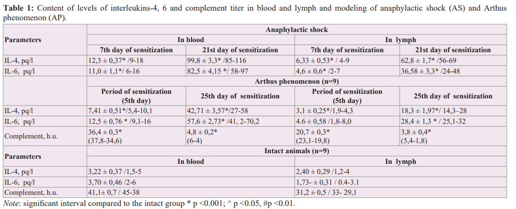
As a result of the study, it was established that both at the stage of sensitization and the resolving stage of anaphylactic shock, the level of IL-4 increases, while IL-6 decreases. On the other hand, in the Arthus phenomenon, IL-4 decreases, and IL-6 increases. Thus, at the stage of sensitization of anaphylactic shock (day 7), the level of IL-4 and IL-6 synthesized by Th3-lymphocytes increases both in anaphylactic (anaphylactic shock) and local (Arthus phenomenon) allergic reactions. This increase was especially pronounced in animals with reproduced anaphylactic shock on day 21 of sensitization, i.e. at the stage of shock. So, if the level of IL-4 during the sensitization period of anaphylactic shock (on the 7th day) increased 3.8 times compared with the intact parameter (p <0.001), but at the stage of anaphylactic shock, it was 31 times higher (p <0.001 ) compared with the parameters of intact animals. If the level of IL-6 in the blood on the 7th day of sensitization increased 3 times (p <0.001) compared with the intact group (p <0.001), but during the period of anaphylactic shock it was 22.3 times higher (p <0.001) than cytokine indices in intact animals.
During the Arthus phenomenon, related to immunocomplex reactions, the levels of immunoglobulins changed as follows. The cytokine parameters of rabbits, although they were increased compared to those of intact, were lower than in animals with reproduced anaphylactic shock. So, if during the period of sensitization (5th day) of the Artyus phenomenon, compared with the intact parameters, the level of IL-4 in the blood was increased by
2.3 times (p <0.001), and the level of IL-6 by 3.4 times (p < 0.001), but in rabbits on the 25th day i.e. during the Artyus phenomenon, compared with intact parameters, the level of IL-4 was 13.3 times lower (p <0.001), and IL-6 was 15.6 times (p <0.001) higher.
Changes in cytokine parameters in lymph, compared with blood, were somewhat weakly expressed. So, if during the sensitization period of anaphylactic shock (7th day), the level of IL-4 and IL-6 compared to intact indicators was 2.6 (p<0.001) and 2.7 (p<0.001) times higher, respectively, but during anaphylactic shock, the level of IL-4 and IL-6 in lymph was 26.1 and 21.3 times higher than intact parameters (p<0.001). If during the Artyus phenomenon on the 5th day of sensitization, the level of IL-4 in the lymph compared with the intact group increased 1.7 times (p <0.05), and IL-6 2.7 times (p <0.001), but on the 25th day i.e. during the Arthus phenomenon, the level of IL-4 and IL-6 were, respectively, 10.7 (p <0.001) and 11.8 (p <0.001) times higher than intact parameters.

Discussions
Akcakaya N., Hashimoto S. et al. indicated an increase in the levels of IL-4 and IgE in the blood serum of patients with atopic bronchial asthma [7]. In sepsis (including the induced by E. coli LPS), IL8 production is regulated by C5a and TLR agonists. The combined inhibition of C5 (or C3) and the TLR coreceptor (CD14) significantly attenuates inflammation and thrombus formation in coli-induced sepsis. During the onset of early atherosclerotic atheromas, cholesterol crystals induce the activation of HCS- dependent inflammasomes with the participation of C1q and C5a, as well as the release of cytokines. The combination of C5a and TNF acts as a potential primer for cholesterol-induced IL1-beta release by increasing IL1-beta transcripts [8].
In our studies, the level of IL-4 increased in the blood and lymph both during the sensitization and the resolving period of anaphylactic shock, and the complement titer decreased both during the sensitization and the shock period. In animals with the reproduced Artyus phenomenon, although during the period of sensitization the level of IL-4 increased slightly, during the period of the Arthus phenomenon the level of this immune parameter significantly decreased. And the level of IL-6 increased at the stage of sensitization and reached its maximum during the period of the Arthus phenomenon. The titer of the complement decreased both at the stage of sensitization and during the period of the Arthus phenomenon, which indicates that a decrease in the activity of complement leads to an increase in the CIC in the blood and lymph. During the shock period (on the 21st day of sensitization), sneezing and involuntary urination were noted in some animals, and loss of balance in others. After suffering anaphylactic shock, 2 animals died, and therefore, blood for the study was taken from only 7 animals.
Toshitani A et al. studied the production of IL-6 in patients with atopic dermatitis, psoriasis, and healthy individuals [9]. There was a significant increase in the activity of IL-6 in patients with atopic dermatitis, in comparison with healthy people and patients with psoriasis. In animals with reproduced Arthus phenomenon, our studies also showed a significant increase in IL-6 activity in comparison with intact animals at the resolving stage. At the site of the antigen injection, hyperemia was first noted, and then the development of infiltration. Further, during the formation of a focus of hyperergic inflammation (20-25th days of sensitization) in experimental animals, tissue hyperemia surrounded by necrosis was noted at the injection site. In some animals, necrosis occupied a large area of the site, in others, only hyperemia without necrosis was noted. In all animals, an increase in blood coagulability was noted, both during the period of sensitization and shock.
The Results of Our Research Have Shown:
- In atopic reactions, the level of IL-4 in the blood and lymph increases, the titer of complement decreases at the stage of sensitization, which is not determined at the stage of anaphylactic shock.
- And in the case of immunocomplex reactions, on the contrary, the level of IL-6 increases, and IL-4 decreases. IL-6 has little effect on Ig E concentration.
- The titer of complement decreases in both experimental allergic reactions.
- The increase in the level of IL-4 and IL-6 in the blood is more significant than in the lymph.
References
- Sudowe S, Arps V, Vogel T, et al. The role of interleukin-4 in the regulation of sequential isotype switches from immunoglobulin G1 to immunoglobulin E antibody production. Scand J Immunol. 2000; 51: 461-471.
- Prosekova EV, Derkach VV, Shestovskaya TN, et al. Cytokine profile and dynamics of IgE synthesis in allergic diseases in children. Immunology. 2010; 3: 140-143.
- Sarrias MR, Farnós M, Mota R, et al. CD6 binds to pathogen- associated molecular patterns and protects from LPS-induced septic shock. Proc Natl Acad Sci USA. 2007; 104: 11724- 11729.
- Voronina MS, Shilkina NN, Vinogradov A, et al. Interleukins-4, 6, 8 in the pathogenesis of rheumatoid arthritis and its complications. Cytokines and inflammation. 2014; 46-48.
- Gushchin IS. IgE-mediated hypersensitivity as a response to impaired tissue barrier function. Immunology. 2015; 36: 45-52.
- Kornienko AA, Kulikovskiy NN, Sorokatyy AE, et al. Catheterization of the thoracic duct in the Experiment. Actual issues of topographic anatomy and surgical surgery. 1977; 1: 22-26.
- Akcakaya N, Sozer V, Corugras H, et al. A preliminary study on IL-4 levels in xtrinsic atopic asthmatic-children. Turk J Pediatr. 1994; 36: 105-110.
- Samstad EO, Niyonzima N, Nymo S, et al. Cholesterol crystals induce complement-dependent inflammasome activation and cytokine release. J Immunol. 2014; 192: 2837-2845.
- Hashimoto S, Amemiya E, Tomita Y, et al. Elevation of soluble IL-2 receptor and IL-4, and nonelevation of IFN-gamma in sera from patients with allergic asthma. Ann Allergy. 1993; 71: 455-458.